drperiklisperikleous.co.uk
  • Home
  • About me
  • Expertise
  • Videos
  • Bronchoscopy
  • Lung Cancer
    • Diagnosis to Treatment
    • Information about surgery
  • Volume Reduction
    • Diagnosis to Treatment
    • Information about surgery
  • Chest Wall
    • Pectus Diagnosis
    • Pectus Surgery
  • Research
  • Teaching
  • Blog
  • Contact
  • More
    • Home
    • About me
    • Expertise
    • Videos
    • Bronchoscopy
    • Lung Cancer
      • Diagnosis to Treatment
      • Information about surgery
    • Volume Reduction
      • Diagnosis to Treatment
      • Information about surgery
    • Chest Wall
      • Pectus Diagnosis
      • Pectus Surgery
    • Research
    • Teaching
    • Blog
    • Contact
  • Home
  • About me
  • Expertise
  • Videos
  • Bronchoscopy
  • Lung Cancer
    • Diagnosis to Treatment
    • Information about surgery
  • Volume Reduction
    • Diagnosis to Treatment
    • Information about surgery
  • Chest Wall
    • Pectus Diagnosis
    • Pectus Surgery
  • Research
  • Teaching
  • Blog
  • Contact

Diagnosis and Assessment

Chest wall deformities

The chest wall is formed by the sternum, the ribs and the costal cartilages that join them. When these cartilages grow unevenly, the shape of the chest changes. This is common, and in most cases it occurs on its own without any associated disease.


There are three main patterns:


Pectus excavatum, or funnel chest, where the sternum is pushed inwards to create a central depression. It is the most common form, affecting around 1 in 400 people, and is about four times more frequent in men.


Pectus carinatum, or pigeon chest, where the sternum protrudes outwards. It is less common than excavatum and tends to become noticeable later in adolescence.


Pectus arcuatum, an uncommon mixed pattern in which the upper sternum protrudes with a depression below it. It does not behave like ordinary excavatum and is not always suited to the same operation, so it needs to be assessed on its own terms.
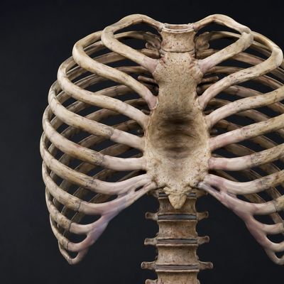
Human ribcage and spine anatomy detail.

How it presents

Most chest wall deformities become apparent during the adolescent growth spurt, usually between the ages of 11 and 16, when the chest grows quickly and the shape becomes more pronounced. Some are noticed in early childhood and change very little. Others progress over a year or two and then stabilise once growth is complete.


Patients come forward for different reasons. Some notice breathlessness on exertion, or find they tire more quickly than others of similar fitness. Some describe chest discomfort. Many have no physical symptoms at all and are concerned by the appearance of the chest.


Concern about appearance is a recognised consequence of chest wall deformity and a common reason for referral. Under current NHS commissioning arrangements it does not, in itself, meet the threshold for surgical funding. Alternative options are outlined below.

Seeing your GP

Your GP will examine the chest and ask about exercise tolerance, breathlessness and chest pain, along with any family history of a similar chest shape — which is present in around 40% of cases.


It is also worth considering associated conditions. A small proportion of patients with pectus excavatum have an underlying connective tissue disorder such as Marfan or Ehlers-Danlos syndrome, and scoliosis is more common than in the general population.

Learn More

Pectus Excavatum

How NHS treatment is decided

Surgery for pectus excavatum is not routinely commissioned by the NHS. It is funded case by case, and every patient must be discussed at a national pectus multidisciplinary team meeting before surgery can be offered.


To be discussed, a patient needs to meet defined criteria:


  • A Haller index of 3.25 or above on CT, and


  • Evidence of cardiac compression on echocardiogram, or on cardiac MRI where the echocardiogram is not definitive, and/or physiological restriction attributable to the deformity on cardiopulmonary exercise testing


If the MDT agrees the criteria are met, surgery can be offered on the NHS, and it is carried out in only a small number of designated centres.


Investigations are completed before the case is presented. Patients do not attend the meeting. The outcome and subsequent plan are discussed afterwards.

Investigations

CT thorax

Cardiopulmonary exercise testing

Pulmonary function tests

Provides cross-sectional imaging and allows measurement of the Haller index, calculated as the transverse diameter of the chest divided by the anteroposterior distance between the sternum and the vertebral column at the point of maximum depression. 

Pulmonary function tests

Cardiopulmonary exercise testing

Pulmonary function tests

Measure lung volumes and airflow. In pectus excavatum, resting values are frequently within normal limits despite significant exertional symptoms, which is why exercise testing is often more informative.

Cardiopulmonary exercise testing

Cardiopulmonary exercise testing

Cardiopulmonary exercise testing

Measures ventilation, gas exchange and cardiac response during graded exercise. Limitation in pectus excavatum characteristically appears under load rather than at rest, and CPET frequently accounts for the discrepancy between normal resting investigations and exertional symptoms.

Echocardiography

Echocardiography

Cardiopulmonary exercise testing

Ultrasound assessment of cardiac structure and function. It demonstrates whether the sternum is compressing the heart, most commonly the right ventricle, and whether filling or function is affected. 

Cardiac MRI

Echocardiography

Cardiac MRI

MRI gives unobstructed imaging and a direct, reliable measure of right ventricular function. Sequences can be timed to demonstrate whether compression varies through the respiratory cycle.

If the criteria are not met

Understanding the threshold

Understanding the threshold

Understanding the threshold

The threshold is deliberately high, and a proportion of patients with a visible and troubling deformity will not meet it. That reflects how the funding criteria are drawn rather than how significant the deformity is to the person living with it.

Vacuum bell therapy

Understanding the threshold

Understanding the threshold

A suction device worn on the chest for a period each day, which gradually lifts the sternum. It works best in younger patients with a flexible, symmetrical and relatively mild deformity, and it requires consistent use over months to years. Most patients buy the device themselves; some NHS services help with fitting and monitoring.

Private treatment

Understanding the threshold

Private treatment

Where NHS funding is not available, surgery and vacuum bell therapy can still be obtained privately, as the commissioning criteria do not apply outside the NHS. Patients wishing to explore this can approach private healthcare providers directly. Many insurance policies classify pectus correction as cosmetic and exclude it.

Pectus Carinatum

The pathway is different

First-line treatment is a compressive brace, worn for several hours a day over 12 to 24 months. It works well in growing adolescents whose chest is still flexible, and within the NHS it is arranged through orthotics. Bracing is considerably more effective in carinatum than in excavatum, so it is genuinely the treatment of choice rather than a fallback.


Surgery for carinatum is not commissioned by the NHS. Assessment is available on the NHS and usually consists of CT and cardiopulmonary exercise testing, and in selected cases of extreme or painful deformity surgery may still be considered. For most patients, however, surgical correction of carinatum is a private procedure.

Research

I am the principal investigator at St George's for the RESTORE trial, a UK study in pectus excavatum surgery. Recruitment has now closed and results are awaited. Taking part in national research is part of how this service stays current, and it is one reason patients here are assessed against contemporary standards.

Find out more

Dr Periklis Perikleous, Consultant in Thoracic Surgery at St George's University Hospitals NHS Foundation Trust in London, United Kingdom


With practicing privileges at Spire St Anthony's Hospital in London, United Kingdom


Copyright © 2026 drperiklisperikleous.co.uk

This website uses cookies.

We use cookies to analyze website traffic and optimize your website experience. By accepting our use of cookies, your data will be aggregated with all other user data.

DeclineAccept